Back Muscles Anatomy Chart : Intermediate And Deep Muscles Of The Back Anatomy Tutorial Youtube - Buy back anatomy chart at amazon.. This rotator cuff muscle helps with the raising and lowering of the upper arm.; Facebook twitter google+ linkedin stumbleupon tumblr pinterest reddit vkontakte share via email print. Other muscles that aid in shoulder movement include: The intermediate layer contains the erector spinae muscles, whose many functions include the extension and lateral flexion of the spine, head and neck. While muscles like the gluteals (in the thighs) are used any time we walk or climb a step, deep back muscles and abdominal muscles are usually not actively engaged during everyday activity.
Your clients will thank you for it! Related posts of muscles of the lower back and hip diagram back muscle chart. All about the back muscles the back anatomy includes the latissimus dorsi, trapezius, erector spinae, rhomboid, and the teres major. A large muscle group in the shoulder, neck and upper back that pulls the head and shoulders backward. Build wide lats with this back building exercise.
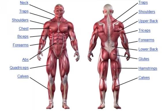
Female Torso Musculature Labelled Back Muscles Anatomy Anatomy Of Muscles Hip And Lower Back Human Muscle Anatomy Body Anatomy Shoulder Muscle Anatomy from i.pinimg.com Related posts of back muscles chart muscle anatomy fitness. Both the deltoid and the trapezius are firmly attached to the spine of the scapula. The muscles of the back are a group of strong, paired muscles that lie on the posterior aspect of the trunk they provide movements of the spine, stability to the trunk, as well as the coordination between the movements of the limbs and the back muscles are divided into two large groups: Other muscles are small and cover much less space. The superficial group, the deep group, and the intermediate group. The back's muscles start at the top of the back (named the cervical vertebrae) and go to the tailbone (also named the coccyx). The muscles of the lower back help stabilize, rotate, flex, and extend the spinal column, which is a bony tower of 24 vertebrae that gives the body structure and houses the spinal cord. Lower back muscle anatomy includes the multifidus, longissimus, spinalis, and quadratus lumborum.
The muscles of the lower back help stabilize, rotate, flex, and extend the spinal column, which is a bony tower of 24 vertebrae that gives the body structure and houses the spinal cord.
For more anatomy content please follow us and visit our website: The deltoid, teres major, teres minor, infraspinatus, supraspinatus (not shown) and subscapularis muscles (not shown) all extend from the scapula to the humerus and act on the shoulder joint. Back muscle chart 12 photos of the back muscle chart back muscle diagram human body, back muscle diagram pain, back muscle groups diagram, back muscle workout diagram, lower back muscle chart, human muscles, back muscle diagram human body, back muscle diagram pain, back muscle groups diagram, back muscle workout. Brings shoulders and arms back to body. While muscles like the gluteals (in the thighs) are used any time we walk or climb a step, deep back muscles and abdominal muscles are usually not actively engaged during everyday activity. The latissimus dorsi muscles (also known as the lats) are the largest muscles of the back. They lift and tilt head and lift or steady the shoulders. Anatomy chart courtesy of fcit. We'll start with the two largest muscles of the back musculature. Moves humerus (arm) to chest. Human anatomy chart find the best weight lifting exercises that target each muscle or groups of muscles. Back muscle diagram back muscles big back big back muscles big lats bodybuilding secrets major back muscles. The trapezius and latissimus dorsi muscles connect the upper limb to the vertebral column.
For more anatomy content please follow us and visit our website: This large muscle in the back. The human back, also called the dorsum, is the large posterior area of the human body, rising from the top of the buttocks to the back of the neck. We are pleased to provide you with the picture named anatomy of back muscles diagram.we hope this picture anatomy of back muscles diagram can help you study and research. Knowledge of the structures in your lumbar spine can also help you communicate with your doctor about lower back problems.
Muscle Anatomy Human Anatomy Chart from www.kingofthegym.com The latissimus dorsi muscles (also known as the lats) are the largest muscles of the back. Lower back muscle anatomy includes the multifidus, longissimus, spinalis, and quadratus lumborum. The human back, also called the dorsum, is the large posterior area of the human body, rising from the top of the buttocks to the back of the neck. Facebook twitter google+ linkedin stumbleupon tumblr pinterest reddit vkontakte share via email print. All about the back muscles the back anatomy includes the latissimus dorsi, trapezius, erector spinae, rhomboid, and the teres major. Certain back muscles extend to other areas, like the shoulders, upper arms, and thighs. Muscle anatomy fitness 12 photos of the muscle anatomy fitness muscle anatomy and workout, muscle anatomy fitness, muscle anatomy workout, muscle anatomy workout chart, muscle and fitness bodybuilders anatomy chart, human muscles, muscle anatomy and workout, muscle anatomy fitness. We are pleased to provide you with the picture named anatomy of back muscles diagram.we hope this picture anatomy of back muscles diagram can help you study and research.
1) make midline incision along spines of vertebrae 2) extend from
The latissimus dorsi muscles (also known as the lats) are the largest muscles of the back. Related posts of back muscles chart muscle anatomy fitness. They lift and tilt head and lift or steady the shoulders. Muscle anatomy fitness 12 photos of the muscle anatomy fitness muscle anatomy and workout, muscle anatomy fitness, muscle anatomy workout, muscle anatomy workout chart, muscle and fitness bodybuilders anatomy chart, human muscles, muscle anatomy and workout, muscle anatomy fitness. See how exercise helps the back. While muscles like the gluteals (in the thighs) are used any time we walk or climb a step, deep back muscles and abdominal muscles are usually not actively engaged during everyday activity. Facebook twitter google+ linkedin stumbleupon tumblr pinterest reddit vkontakte share via email print. The two trapezius muscles extend from the backbone and base of the skull, across the back and shoulders to join the scapula and the clavicle. The pelvic floor muscles also help increase this pressure, which provides stability to the spine and trunk. 1) make midline incision along spines of vertebrae 2) extend from The intermediate layer contains the erector spinae muscles, whose many functions include the extension and lateral flexion of the spine, head and neck. The back's muscles start at the top of the back (named the cervical vertebrae) and go to the tailbone (also named the coccyx). The muscles of the chest and upper back occupy the thoracic region of the body inferior to the neck and superior to the abdominal region and include the muscles of the shoulders.
1) make midline incision along spines of vertebrae 2) extend from The back's muscles start at the top of the back (named the cervical vertebrae) and go to the tailbone (also named the coccyx). A large muscle group in the shoulder, neck and upper back that pulls the head and shoulders backward. On this page, you'll learn about each of these muscles, their locations and functional anatomy. All about the back muscles the back anatomy includes the latissimus dorsi, trapezius, erector spinae, rhomboid, and the teres major.
Paraspinal Muscles Anatomy Paraspinal Muscles Anatomy Human Anatomy Library Photo Paraspinal Muscles A Shoulder Muscle Anatomy Shoulder Anatomy Muscle Diagram from i.pinimg.com Brings shoulders and arms back to body. This large muscle in the back. The pelvic floor muscles also help increase this pressure, which provides stability to the spine and trunk. For more anatomy content please follow us and visit our website: Buy back anatomy chart at amazon. Related posts of muscles of the lower back and hip diagram back muscle chart. Other muscles are small and cover much less space. They lift and tilt head and lift or steady the shoulders.
Anatomy chart courtesy of fcit.
Superficial back muscles, intermediate back muscles and intrinsic back muscles.the intrinsic muscles are named as such because their embryological development begins in the back, oppose to the superficial and intermediate back muscles which develop elsewhere and are therefore classed as extrinsic muscles. Human anatomy chart find the best weight lifting exercises that target each muscle or groups of muscles. See how exercise helps the back. Free shipping on qualified orders. Related posts of muscles of the lower back and hip diagram back muscle chart. The two trapezius muscles extend from the backbone and base of the skull, across the back and shoulders to join the scapula and the clavicle. Muscle charts of the human body. Muscles found in the superficial group include rhomboid major, rhomboid minor, levator scapulae, trapezius, latissimus dorsi. We'll start with the two largest muscles of the back musculature. Some of these muscles are quite large and cover broad areas. The trapezius and latissimus dorsi muscles connect the upper limb to the vertebral column. This large muscle in the back. Claim your free copy of the client back care guide today.
Back muscles, like any other muscle in the body, require adequate exercise to maintain strength and tone back muscles chart. Muscles found in the superficial group include rhomboid major, rhomboid minor, levator scapulae, trapezius, latissimus dorsi.


0 Comments:
Post a Comment